ABOUT US
Throughout our 30+ year history, LICORbio has developed revolutionary fluorescence imaging technologies to address the growing needs of the global scientific community. Today LICORbio offers complete, integrated solutions for quantitative protein research, immunocytochemistry, virology, small animal imaging, and 2D and 3D cell culture imaging including z-stacks, morphology analysis, organoids, spheroids, and microphysiological systems.
The LICORbio Atlas™ Imaging System is the first in a new line of 3D cell culture imaging systems. Created for the demands of translational research, the LICORbio Atlas Imager delivers fast, precise 2D and 3D imaging without the need for stitching, intensity correction, or other time-consuming post-processing steps. It delivers reliable, quantifiable results in less time, so researchers can keep their focus where it belongs: on their next discovery.
The unique line-scanning technology of the LICORbio Atlas Imager enables whole-plate imaging in less than one minute, as well as higher-resolution, single-well scanning. It can also image through entire 3D samples in 0.01 mm increments to create stunning z-stacks and 2D projections of organoids, spheroids, and organ-on-a-chip systems. With over 30 imaging channels spanning UV to NIR, plus luminescence, brightfield, and epi-illumination, the LICORbio Atlas Imager simplifies workflows, eliminates inefficiencies, and captures deep spatial insights in a fraction of the time—and at a fraction of the cost—of competitive solutions.
With over 120,000 citations in peer-reviewed publications, industry-leading imaging systems, powerful analysis software, affordable reagents, and world-class scientific support, LICORbio empowers researchers around the world to answer difficult questions, make groundbreaking discoveries, and improve countless facets of human health. LICORbio is an ISO 9001:2015-certified company.
FEATURED ARTICLES
-
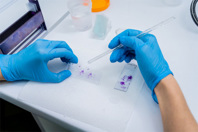
See how total protein staining offers a robust approach to western blot normalization to minimize variability and improve quantification accuracy, which supports reproducible protein expression.
-
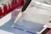
Review this step-by-step guidance for fluorescent western blot detection, including membrane prep and antibody handling, to improve signal clarity and consistency across imaging platforms.
-
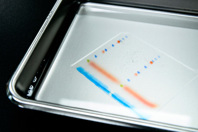
Discover how to determine the linear range for western blot quantification using total protein stains or housekeeping proteins to ensure accurate normalization and avoid saturation effects.
-
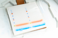
Gain insight into how to choose and validate normalization strategies for western blots, avoid common pitfalls, and ensure reproducible results using proteins tailored to your experimental context.
-
Learn how to design, normalize, and interpret replicate western blot experiments to ensure reproducible protein quantification with fold change calculations, CV analysis, and visualization strategies.
CONTACT INFORMATION
LICORbio
4647 Superior Street
Lincoln, NE 68504
UNITED STATES
Phone: 402-467-0800
Contact: Katie Schaepe

