
What Is EPR Spectroscopy? Principles, Spin Labeling, And DEER In Proteins
By Austin Gamble Jarvi
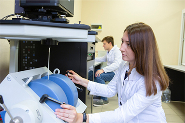
Understanding biomolecular function requires capturing full structural ensembles rather than relying solely on static snapshots. Electron Paramagnetic Resonance (EPR) spectroscopy addresses this need by detecting unpaired electron spins through site-directed spin labeling, delivering highly selective measurements without background interference.
By isolating dipolar interactions, methods like Double Electron-Electron Resonance (DEER) measure interspin distances across 1.5 to 8 nm, revealing distance probability distributions that report directly on conformational flexibility and heterogeneous states. This dynamic insight complements traditional techniques like Cryo-EM, X-ray crystallography, and NMR, resolving structural ambiguity in complex membrane proteins, intrinsically disordered proteins, and metalloproteins. Review the full article to evaluate how integrating EPR spectroscopy refines structural characterization and illuminates dynamic biomolecular mechanisms.
Get unlimited access to:




Enter your credentials below to log in. Not yet a member of Bioprocess Online? Subscribe today.