
Universal Imaging Upgrades MetaMorph for Six-Dimensional Imaging
Imagine that you want to acquire DAPI and FITC wavelength images and also want to acquire both wavelengths through ten different focus "Z" planes. And then you also want to acquire those images over time, say every two minutes for an hour. All this can be done in MetaMorph without having to "program" a journal or macro. The "Multi-Dimensional Acquisition" dialog guides you through the acquisition process no matter what combination of image dimensions your research requires. New users with little or no training could be running complex acquisition paradigms within minutes.
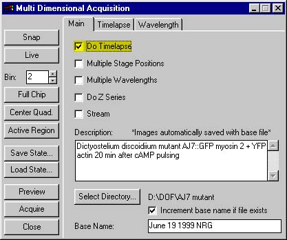
Once you have acquired a sophisticated set of multi-dimensional images, you can use MetaMorph's new imaging tools to analyze, process, and review your images. In the example above, you acquired 600 images (2 wavelengths X 10 planes X 30 time points = 600 images). Loading one image at a time for processing or making measurements is tedious and time-consuming. MetaMorph's "Review Multi-Dimensional Data" dialog allows you to readily view and process all or parts of the image data set at your discretion. You can make movies, color-combine images, measure images, and do 3D reconstruction—it makes no difference how many images you have. With the new MetaMorph tools, it's as easy to process six hundred images as it is to process one.
"Movie Chemistry"
Mark Davis, professor of Microbiology and Immunology at Stanford University, is doing what he calls "movie chemistry" in his research on T-cell activation. Having exhausted what conventional biochemical technology on isolated receptor molecules could tell him about the process, Davis turned to live cell imaging to get at the events that take place when T-cell receptors become engaged.
One approach Davis wished to take was to construct three-dimensional images of activated T-cells using a combination of fluorescent dyes—one to monitor the T-cell receptors on the surface, and one to measure calcium flux, a benchmark of T-cell activation. However, he discovered that conventional image analysis set-ups couldn't capture the events he was after fast enough. Acquiring the images took so much time that the cells moved while the information was being acquired, which caused smearing of the image.
Using UIC's prototype 6-D technology and a piezo-electric focusing device to move the objective and speed up the z-series, Davis could cut the time required for an acquisition cycle from 30 seconds to 2.5–3 seconds. With the new procedure, he captured 20 to 25 16-bit images, 650 X 500 in size with 50 to 100 millisecond exposures and one to 1.5 micron steps, creating a 3D "snapshot" at each time point and z position. With this rate of acquisition, Davis was able to look at the dynamic events in live moving T cells, revealing a balletic cycle of movement of T-cell receptors, as they coalesced into a bull's-eye and dissipated repeatedly, all the while driving calcium in and out of the cell.
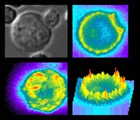
This montage of GFP-labeled cells demonstrates high-speed 3D-reconstruction, or z-streaming.
For additional thoughts on Multi-Dimensional Imaging, check out "Visual Reality: Using Computer Reconstruction and Animation to Magnify the Microscopist's Perception" in Molecular Biology of the Cell (October 1999) Vol. 10: 3061-3065.
For more information: Matt Harrington, Marketing Communications Mgr., Universal Imaging Corporation, 502 Brandywine Pkwy., West Chester, PA 19380. Tel: 610-344-9410, ext. 221.