
TissueInformatics develops bioinformatics approaches to analyzing cell and tissue structures
Company provides image processing and data management tools for tissue-derived information
By Tom Hollon
Tissue informatics, the application of bioinformatics to histology, fills an important gap left open in the movement to assemble giant databases that capture every last detail of the human body's composition. The foundation for 21st century biology rests upon the vast Human Genome Project databases. Anatomy in our new century will rely upon the Visible Human databases under development at the National Library of Medicine from digitized photographs of millimeter-thin slices of frozen male and female cadavers. And in between molecular biology and anatomy lies another field ripe for bioinformatics—the study of cell and tissue structure.
In 1997, Peter C. Johnson, then a physician at the University of Pittsburgh, realized that bioinformatics could be applied to the microscopic analysis of tissue samples, the bread-and-butter technique of laboratory pathology. Digital imaging techniques could make cell and tissue structure analysis completely quantitative and automated. And by combining image analysis of cells and tissues with gene expression data, scientists mining the genome could take away deeper insights about how genes influence disease. Inspired by this vision, Johnson founded Pittsburgh-based TissueInformatics, Inc.. His young company applies its new brand of bioinformatics in the service of tissue engineering and drug discovery.
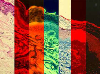
The information gleaned from tissue depends upon the different stains, light wavelengths, and protein and gene expression techniques used. Automated analysis software quantifies all cell components that detection techniques make visible.
For one tissue engineering company, TissueInformatics develops software that will automate quality control of its engineered skin tissue products. "Skin tissues must be shipped to waiting hospitals within hours of their creation to maximize their useful lifespan," says Johnson; "Quality control must not only be accurate, but rapid."
"Moreover," he adds, "it must detect changes in product quality early."
Using microscope-mounted robotics, the software automatically captures images of engineered skin mounted on microscope slides and analyzes their health. That includes not only individual cells, but the skin as a whole. Analyzing cell numbers and distribution, cell density and organization, cell shape and thickness of layers, the software certifies skin structure as normal or abnormal. Company scientists will use the software to reduce lot-to-lot product variation, and study the images for ways to improve skin engineering.
The watchword for TissueInformatics imaging analysis is versatility; the company's software can recognize histological stains and cell structures (nuclei, for instance), or work with lighting techniques like fluorescence or expression techniques like in situ hybridization. If the eye can see it, machine vision software can analyze and quantify it. Even what the eye cannot see—a technique called spectral imaging extends analysis to ultraviolet and infrared wavelengths.
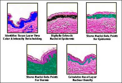
TissueInformatics analyzes tissue structure by quantifying the locations of cells and cellular components. The data can be queried for any information visible with the tissue. Here a skin sample is analyzed.
TissueInformatics also earns its keep in drug companies, providing cell and tissue analysis services for "corporate pathologists," as Johnson calls them, who spend their microscope hours examining the same types of cells over and over, hunting signs of benefit or toxicity in tissues exposed to experimental drugs. Drug companies build virtual tissue banks of these images, annotated with analysis results, for further study. By 2002, they'll be able to compare their images online against reference images of healthy and diseased tissues in TissueInformatics' own database.
Johnson is building his company through software products like a Dermf(x), which characterizes and quantifies skin features. Besides skin engineering, it's useful for transdermal drug delivery projects and development of wound healing drugs and cosmetics.
He also sees a big future in databases combining gene and protein expression analysis with tissue informatics. Here will be a superb way, he declares, for scientists to correlate molecular activity to cellular activity, yielding richer understanding of disease and drug effects. He's building his company to lead the way.
For more information, contact: Peter C. Johnson, President and CEO, TissueInformatics, 711 Bingham St., Suite 202, Pittsburgh, PA 15203. Tel: 412-488-1100. Fax: 412-488-6172.
About the author: Tom Hollon is a science writer and editor based in Rockville, Maryland. He was the founding editor of the journal Modern Drug Discovery. Prior to that, Tom conducted research at the National Institutes of Health, the Pasteur Institute, and the University of Washington. He can be reached at thollon@starpower.net.