
Penn scientists create probe for amyloid plaque that crosses the blood-brain barrier

Their work—which represents the first time that amyloid deposits have been visualized in live cells—sets the stage for the refinement of this compound so it may be used to diagnose and monitor the treatment of patients with Alzheimer's disease. At the present time, diagnostic confirmation and visualization of the plaque burden in the brains of Alzheimer's patients can occur only after death, during postmortem autopsy.
The design and synthesis of BSB was the work of Hank Kung, a co-author of the study, who is known for creating other molecules that have become essential diagnostic brain-imaging probes now in use worldwide. The researchers first established that BSB could enter cells by treating cultured human cells with the compound. They further showed that brain cells could take up BSB by injecting the compound into the brains of non-diseased mice. They turned next to a transgenic mice modeled for Alzheimer's disease. Injecting BSB directly into the hippocampi of transgenic mice revealed, upon post-mortem analysis, clear and distinctive visual staining of the amyloid plaques.
"Our next step was the Holy Grail," remembers Lee, whose team still had to prove that their new compound was strong enough, small enough, and soluble enough to efficiently pass through the blood-brain barrier. To that end, BSB was injected into the veins of transgenic mice. Again, post-mortem studies of the animals' brains showed clearly-discernible plaques. In addition, the investigators found that BSB remained bound to amyloid plaques for at least 18 hours. "We demonstrated unequivocably that the compound can go through the blood-brain barrier and bind to amyloid," says Lee.
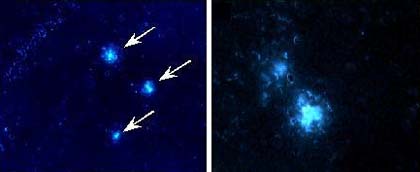
Intravenous injection of BSB results in abundant labeling of senile plaques in transgenic mice (Tg2576). Direct examination showed prominent BSB staining of plaques in the hippocampus (left) and entorhinal cortex (right). Arrows indicate the position of staining with anti-amyloid b peptide specific antibody (data not shown).
"This is definitely proof of concept," says Lee, professor of pathology and laboratory medicine at Penn and lead author of the study that appears in the June 20 issue of Proceedings of the National Academy of Science. "We have demonstrated for the first time that a flourogenic probe can cross the blood-brain barrier and bind to amyloid plaques in the brains of transgenic mice that develop such plaques. This is an essential first step for the development of an antemortem diagnostic for Alzheimer's disease."
"This research is an enormously important step toward developing an imaging method that could pinpoint the telltale signs of plaque development associated with Alzheimer's disease in a living brain," echoes Marcelle Morrison-Bogorad, associate director of neuroscience and neuropsychology of aging at the National Institute on Aging (Bethesda, MD), which provided the major funding for the study.
"This tool could help clinicians peer into a person's brain and monitor amyloid levels in response to treatment. We definitely need something like this to advance the diagnosis and treatment of dementia."
Lee and her colleagues will fine-tune BSB by increasing its solubility or decreasing its molecular weight. According to Dan Skovronsky, first author of the paper, they have already developed a radioactive marker that will allow visualization by PET or SPECT imaging. "The exciting promise of our agent is that, when clinically applied, it will demonstrate efficacy of therapy in treatments designed to inhibit the growth of amyloids," explains Lee.
For more information: Virginia M.-Y. Lee, Center for Neurodegenerative Disease Research, Departments of Pathology and Laboratory Medicine, Radiology, and Pharmacology, University of Pennsylvania School of Medicine, Philadelphia, PA 19104. Email: vmylee@mail.med.upenn.edu.