Not All Plate Counting Technologies Are The Same
By Tim Sandle, Ph.D.

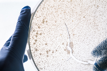
While rapid microbiological methods have advanced, most microbiology laboratory tests remain reliant upon assessing microbial growth on agar plates. Discrepancies with plate counting,1 together with the limitations of human vision,2 have led to regulatory concerns to the extent that the U.S. FDA Q&A document on data integrity recommends “double plate counting” using two independent analysts.3 Issues with plate counting such as the following occur periodically in FDA warning letters:
- Failure to verify counts between operators
- Failure to use suitable equipment to assess colonies (such as not using an appropriate light source)
- Differences that occur in counting between operators
- Counts on plates not matching recorded data
- Loss or damage to plates before data can be captured
An alternative to this practice, and one that meets the regulatory interest in data integrity as well as aiding the economic pressures driving the lean laboratory paradigm, is automated colony counting. Additional advantages that the laboratory can gain from this form of automation include faster times to results, cost control, and digital data capture.
Not all plate counting technologies are identical and some struggle with mixed cultures or growth effects that lead to merged colonies. What is important is the appropriate selection and qualified adoption of automated digital capture colony counters within the pharmaceutical microbiology laboratory. This article considers how such devices can be qualified and how data integrity concerns can be addressed.
Optimal Instruments
The most suitable automated colony counters will be unaffected by the inoculation method, shape, or size of the colony. An effective automated colony counter needs to have good sensitivity for the detection of smaller colonies, particularly in low contrast media, and the ability to capture counts and images using digital technology. In keeping with the automation theme, devices need to be able to transfer data to laboratory information management systems (LIMS). More recent advances include algorithms capable of multi-task learning to both predict the colony centers and conduct colony segmentation.
Colonies can be difficult to count. Some automated devices fail to count more challenging colonies. Less optimal devices have issues such as:4
- Underestimating the numbers of colony forming units (CFUs) on a plate due to overcrowding
- Failing to discriminate colony overlap (i.e., masking), together with interpreting the degree of convexity; colony brightness; variations in colony shape; differences with colony texture
- Failing to assess colonies located near the plate periphery
- Failing to accurately count colonies where increased density occurs
- Inability to assess mixed cultures or variations with colony pigmentation.
Factors affecting the upper countable range include colony size and behavior (such as possible swarming, as is the case with organisms like Proteus mirabilis and some Bacillus species, such as Bacillus cereus), as well as the surface area of the plate. At the lower end of the range, it is important to verify that automated counters can count a single colony.
Key Functionality Of Automated Colony Counters
In terms of functionality, the best automated colony counters should offer:
- Standardized and accurate results. Accuracy is important since colony counting can be affected by numerous parameters related to the physical properties of the colony: size, shape, contrast, and overlapping colonies. To achieve this requires automatic colony separation (for when colonies are positioned close to each other).
- The ability to count colonies within appropriate parameters (such down to 50 microns and measure zones accurately to 0.5 millimeters, within detection limits of 0.1 millimeters).
- Good optical response performance (sufficient control of the background noise, contrast, resolution, etc.) of the image acquisition tools.
- Effective image resolution, file size/data management, sample lighting, and instrument uniformity.
- The ability to visualize white light and fluorescent colonies.
- The ability to differentiate between chromatic and achromatic images and thus deal with both color and clear media.
- The ability to separate aggregated colonies.
- The ability to count the entire plate or sectors of the plate.
- Results obtained within 1 second per plate.
- The display of real-time full-color on-screen images.
- Zoom function for looking at smaller colonies.
- Software to allow for data collection and analysis. Data should ideally be transferrable to a LIMS
The essential elements of automated digital colony counters include a circular dark field illuminator and a camera with a resolution of 3.3 megapixels or higher (many systems have cameras of higher quality); software with appropriate algorithms; and an automated plate holder (with a toolbox to enable communication between the software and the image analyzer). Many of the software algorithms work on the basis of a Bayes classifier. This is a simple probabilistic classifier used to study the geometric properties, such as ratio between major and minor axis of the group, and to verify the number of colonies contained in the group.5
Some types of counters offer greater functionality. For example, some can provide data on the size or shape of the colonies and measure zones of inhibition via software. More sophisticated software can also provide color and size differentiation (allowing microbiologists to distinguish colonies of different hues). Other systems have the functionality to count and calculate concentrations of spiral-plated colonies.
Assessing Data Integrity
As with any other computerized system, the automated colony counter instrument must meet the expectations for laboratory data integrity. When undertaking an assessment, five key data integrity questions are:6
- Is electronic data available?
- Is electronic data reviewed?
- Is meta data (audit trails) reviewed regularly?
- Is there clear segregation of duties?
- Has the system been validated for its intended use?
Validation Of Automated Colony Counters
Validation of automated colony counters is important and necessary for their implementation into the laboratory. To ensure the validity of their data, microbiologists need to establish that their automated colony counting method is as accurate as a precise manual count before they implement any new process into their workflow. This should be conducted across a range, based on the laboratory’s maximum and minimum counting ranges, and include assessments of both pure (as might occur with growth promotion testing) and mixed cultures (as will occur when conducting bioburden determinations). Validation exercises must include multiple assessments.
The acceptance criteria should be based on an acceptance range (the system may slightly undercount and overcount) for both individual plates and for the set of plates assessed. A range of 95%–105% might be appropriate for higher counts, becoming tighter as the population range assessed decreases. This would be based on a comparison of any difference between manually counted and automatic colony counts. For this, an appropriate statistical method for significance should be used, such as a two-tailed t-test for paired samples with a 95% confidence level.7
Validation runs are not always successful. Failures can occur where there are mixed colonies or due to inhomogeneity of the agar thickness, meaning that discrimination is not possible for all areas of the plate. A further weakness is where confluent growth occurs. The light also needs to be suitable. Generally, white light works better than yellow or amber light; however, a blue dark field illumination provides the best discrimination. A further problem that can arise is the presence of debris and other unwanted material, either embedded within or on the surface of the agar.
These weaknesses can sometimes be overcome through reassessing the samples or making adjustments to the instrument. If the weaknesses cannot be addressed, resulting in the acceptance criteria not being met, then the automated colony counter is not suitable for use and instead an alternative instrument needs to be used or manual counting returned to, with each plate checked by a second operator.
References
- Sandle, T. (2018) Automated, Digital Colony Counting: Qualification and Data Integrity, Journal of GXP Compliance, 22 (2): https://www.researchgate.net/publication/324314089_Automated_Digital_Colony_Counting_
Qualification_and_Data_Integrity - Sandle, T. Ready for The Count? Back-To-Basics Review Of Microbial Colony Counting, Journal of GXP Compliance, 24 (1): https://www.researchgate.net/publication/339416178_Ready_for_The_Count_Back-To-Basics_Review_Of_Microbial_Colony_Counting
- FDA. Data Integrity and Compliance With Drug CGMP, Questions and Answers. Guidance for Industry, 2018: https://www.fda.gov/media/119267/download
- Tidswell E. C., Sandle T. Microbiological Test Data - Assuring Data Integrity. PDA J. Pharm. Sci. Technol. 2018, 72 (1), 2–14. https://doi.org/10.5731/pdajpst.`2017.008151
- Deutschmann, S. et al. A Systematic Approach for the Evaluation, Validation, and Implementation of Automated Colony Counting Systems, PDA Journal of Pharmaceutical Science and Technology, 76 (6), 2022: https://journal.pda.org/content/76/6/509.abstract
- Schmitt, S. (2014) Data Integrity, Pharmaceutical Technology Europe, 38 (7). Online edition: https://www.pharmtech.com/data-integrity
- Brugger, S.D., Baumberger, C., Jost, M., Jenni, W., Brugger, U., et al. (2012) Automated Counting of Bacterial Colony Forming Units on Agar Plates. PLOS ONE 7(3): e33695. https://doi.org/10.1371/journal.pone.0033695
About The Author:
 Tim Sandle, Ph.D., is a pharmaceutical professional with wide experience in microbiology and quality assurance. He is the author of more than 30 books relating to pharmaceuticals, healthcare, and life sciences, as well as over 170 peer-reviewed papers and some 500 technical articles. Sandle has presented at over 200 events and he currently works at Bio Products Laboratory Ltd. (BPL), and he is a visiting professor at the University of Manchester and University College London, as well as a consultant to the pharmaceutical industry. Visit his microbiology website at https://www.pharmamicroresources.com.
