
How EPR Spectroscopy Reveals Membrane Protein Structure And Dynamics
By Austin Gamble Jarvi
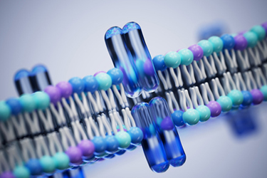
Membrane proteins regulate vital cellular processes and represent a primary class of drug targets, yet their dependence on lipid environments makes them notoriously difficult to characterize. Standard structural biology techniques like X-ray crystallography and cryo-EM capture static snapshots, often missing short-lived, dynamic states critical to understanding biological function and drug binding.
Electron paramagnetic resonance (EPR) spectroscopy bridges this technical gap by measuring nanoscale distance distributions across full conformational ensembles directly in native-like membrane mimetics or live cells. By tracking structural shifts triggered by ligands or environmental changes, EPR delivers crucial insights into complex systems such as GPCRs and ion transporters. Read the full article to discover how integrating EPR spectroscopy into structure-based drug discovery programs resolves complex membrane dynamics and accelerates target characterization.
Get unlimited access to:




Enter your credentials below to log in. Not yet a member of Bioprocess Online? Subscribe today.