
Ferrets Provide Information on Plasticity of the Developing Brain

The ferret offers a unique window into the developing brain, as they are born with relatively undeveloped brains. Capitalizing on this property, Sur and coworkers devised a way to coerce sensory neurons from the retina that normally provide input to the brain's primary visual cortex (VI) to instead innervate the primary auditory cortex (AI). When they did this, they found that the putative auditory brain cells take on the properties of the visual system, both with respect to form and function.
Earlier work from the Sur lab had shown that rewired AI regions are organized like the VI regions to the extent that different neurons are selective for different oriented visual stimuli. In the present work (ref. 2), they looked in detail at higher order features that characterize the normal VI region—the visual orientation columns—whose layout forms the basis of perception of visual stimuli. They found that the AI cells had a pinwheel organization and horizontal connections that are characteristic of the VI region and have no equivalent in the AI region.
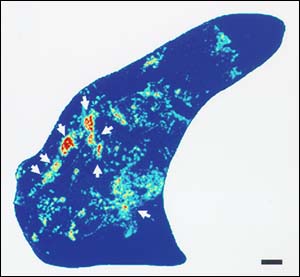
Retinal projections to auditory thalamus in a rewired ferret.
In the second paper (ref. 3), Sur and coworkers show that the animals with rewired brains actually see with their auditory cells. Given the option of receiving a reward from one side following light or the other following sound, the animals responded to the light, which showed that the animals could see light with their auditory cortex.
Taken together these results show that the pattern of activity alone can shape the architecture of the developing cortex. Of this result, Sur told Reuters, "What our work demonstrates is the tremendous capacity for vision to be mediated by new areas of the brain. So if there is blindness that is due to loss of the cortex that is visual, in principle, but not in practice, other areas of the cortex can take over the function of the visual cortex. That is what our work suggests."
References
- Roe, A.W., "Visual projections routed to the auditory pathway in ferrets: receptive fields of visual neurons in the primary auditory cortex," J. Neurosciences 12: 3651-4, 1992.
- Sharma, J, et al., "Induction of visual orientation modules in the auditory cortex," Nature 404: 841-7, 2000.
- Von Melchner, L. et al., "Visual behavior mediated by retinal projections directed to the auditory pathway," Nature 404: 871-866, 2000.
For more information: Mriganka Sur, Department of Brain and Cognitive Science, MIT, 77 Massachusetts Ave., Cambridge, MA 02139-4307. Tel: 617-253-8784. Fax: 617-253-9829. Email: msur@ai.mit.edu.
By Laura DeFrancesco