Determining Residual Bead Count: Application Of Micro-Flow Imaging To CAR T-Cell Manufacturing

Immunotherapy revs up the body’s own natural defenses to fight cancer, veering away from traditional strategies that have, instead, focused on targeting the tumor and tumor cells. All of the enacting immunotherapeutic approaches incite hope, but especially those that uniquely engineer T cells to seek and destroy tumor cells. Chimeric antigen receptor (CAR) T-cell therapies, in particular, have produced booming immune responses and striking clinical outcomes with two such therapies already on the market and 800 clinical trials underway¹.
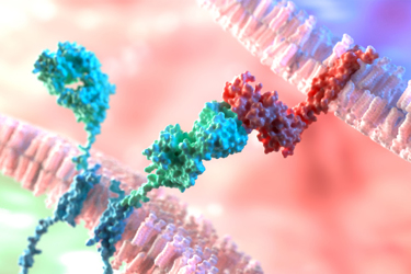
CAR T-cell therapy involves first isolating the patient’s T cells and genetically modifying them to express a CAR on their surface capable of recognizing tumor-associated antigen(s). The engineered cells are then expanded ex vivo to an appropriate therapeutic dose and reinfused into the patient to stimulate an effective T-cell mediated anti-tumor immune response. From a manufacturing perspective, generating personalized batches of inherently complex CAR T-cell products poses real challenges for the biopharmaceutical industry. Regulatory agencies mandate that identity, purity, potency and safety attributes are closely monitored both in-process and for release. Of the process-related factors affecting product purity, residual beads during ex vivo expansion and activation of T cells pose safety and efficacy concerns with regard to triggering an unwanted endogenous immune response in vivo. Thus, compulsory limits are often set for residual bead counts to demonstrate product quality².
Residual bead count is typically determined manually by the naked eye and microscopy. But this approach is highly limited in its ability to accurately discern beads from cells and other potential in-process impurities, resulting in reporting uncertainties that risk regulatory approval. In this application note, you’ll see how automation via image-based Micro-Flow Imaging™ (MFI) technology gets you the quantitative and morphological data you need to have confidence in distinguishing between beads, T cells or other potential contaminants.
Request A Quote
Get unlimited access to:
Enter your credentials below to log in. Not yet a member of Bioprocess Online? Subscribe today.
