Johns Hopkins Team Builds Light-Sensitive Retina Using Human Stem Cells

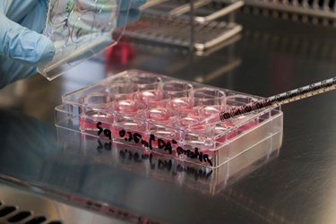
A team of researchers from Johns Hopkins University School of Medicine reports that they have built a 3D complement of human retinal tissue in a lab dish, which contains functional photoreceptor cells that are sensitive to light.
The cells’ sensitivity to light precedes conversion of the stimulus into visual images. “We have basically created a miniature human retina in a dish that not only has the architectural organization of the retina but also has the ability to sense light… [The work] advances opportunities for vision-saving research and may ultimately lead to technologies that restore vision in people with retinal diseases,” says study leader M. Valeria Canto-Soler, an assistant professor of ophthalmology at the Johns Hopkins University School of Medicine.
The team experimented with human induced pluripotent stem cells (iPS), which could eventually lead the way to genetically engineered cells transplants capable of halting or even reversing progressive blindness. The researchers grew the iPS into retinal progenitor cells that make up the light-sensitive retinal tissue found lining the back of the eye.
According to Xiufeng Zhong, postdoctoral researcher in Canto-Soler’s lab, the growth corresponded to the timing and duration of a fetus’ retinal development in the womb. The photoreceptor cells also matured enough to develop outer segments, structures critical to the cells’ function. “We knew that a 3-D cellular structure was necessary if we wanted to reproduce functional characteristics of the retina, but when we began this work, we didn’t think stem cells would be able to build up a retina almost on their own. In our system, somehow the cells knew what to do,” said Professor Canto-Soler.
The researchers tested the dish-grown mini retinas by placing an electrode in a single photoreceptor cell and then giving a pulse of light to the cell, which showed a biochemical pattern reaction similar to photoreceptor behavior in people exposed to light.
The system used by the team allows for the generation of hundreds of mini-retinas at the same time, directly from a patient affected by certain retinal diseases such as retinitis pigmentosa. This offers a novel biological system to directly study retinal disease causes in human tissue instead of depending on animal models. More significantly, the system opens possibilities for personalized treatment for patients.
The team’s work was published online in the journal Nature Communications.
